


Fluoroscopy
A fluorograph is a diagnostic X-ray machine necessary for flow-type, prophylactic X-ray fluorography in order to determine whether a patient has a pathology in the examined area.
Portable (mobile) fluorographs currently available on the market compete well with the stationary models. Such x-rays are of two types – more modern digital ones and nearly forgotten film-based X-rays.
The X-ray film used for capturing images is also used in a film-based fluorograph, in which case a print is obtained on the physical carrier after it is developed. As in all adjacent fields, film is being rapidly replaced by digital fluorography. The film method is already significantly outdated and inefficient in terms of both time, additional, harmful and expensive developing equipment, as well as inconvenience in transferring examination results to colleagues and patients. However, it is worth noting that some doctors who have been performing fluorographic examinations for many years favour traditional methods of obtaining examination results.
You can choose and buy a fluorograph (with an option to pay for it in instalments) for a reasonable price in our online store. When buying, please note the name of the seller and the warranty. Check if there is a valid certificate of conformity and registration certificate.
New digital fluorographs will help effectively monitor the health condition of patients, and will always be needed.
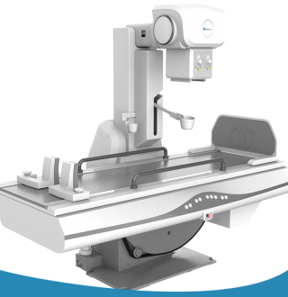
Digital fluorographs
Digital fluorographs are becoming more and more popular in medical institutions, because the examination of thoracic organs for detecting tuberculosis, lung tumours and other bronchial and lung pathologies is a prerequisite for the treatment and follow-up care of all types of patients, and the image quality is several times better compared to outdated technologies.
Unlike film-based machines, digital image processing fluorographs do not require the use of X-ray film to obtain an image – they use a digital receiver for capturing an X-ray image displayed on the monitor screen. This reduces the costs of the examination and frees medical institution from the need to receive supplies in order to be able to carry out the examination.
-
modern Italian and Japanese emitters that emit a minimum “dose” of radiation are more reliable;
-
includes accessories to protect the gonads and thyroid gland;
-
automated doctor and laboratory assistant workstations;
-
provides everything that you need for work – setting imaging modes, setting brightness and contrast, increasing a fragment, determining the area of isolated regions, quick filling out of an examination report.